Science
A pure biological environment, created
and perfected by cells.
What's inside
Proteomics · LC-MS/MSSpecies-verified Pacific salmon
Clean production with cultivated salmon cells instead of farmed salmon sperm, with identical purity, structure, sequence as sperm-derived PDRN.
15,540 peptides (DIA LC-MS/MS)
The peptide fraction — 15,540 distinct sequences identified by mass spectrometry. These short chains span signalling peptides and fragments of larger structural and matricellular proteins; skin cells read many of them as cues rather than bulk material.
5.5 × 10⁹ particles/mL · avg 154.1 nm · 30–200 nm · NTA
Exosomes are how cells talk to one another: nanoscale, lipid-bilayer-bound vesicles that package a cargo of proteins, growth factors, peptides and nucleic acids in one cell and deliver it to another. It is the cargo that carries the signal — the intact vesicle is the messenger that gets it there. In MCC these exosomes are a native fraction of the whole conditioned medium, secreted in biological context rather than isolated, concentrated, or spiked in. Their presence and identity are confirmed by nanoparticle tracking analysis (NTA) and the vesicle markers CD63 and syntenin/SDCBP, verified at a certified independent third-party laboratory.
5,105 proteins (DIA LC-MS/MS)
The structural and matrix proteins a cell builds and secretes — collagens, laminins, fibronectin, fibrillin/elastin-related proteins, matricellular regulators (decorin, thrombospondins), keratins, and thousands more identified by mass spectrometry. This is the scaffold-and-regulator layer of the secretome.
At physiological concentrations
Signalling proteins present at the levels living cells naturally secrete, not super-physiologic single-factor doses. Physiological tuning is the point of a conditioned medium: these factors are read in balance, as cells produce them. A small sample of the numerous growth factors found in MCC is listed below:
- Fibroblast Growth Factor-2 (FGF-2)
- TGF-β family
- Insulin-like Growth Factor (IGF)
- Epidermal Growth Factor (EGF)
- Vascular Endothelial Growth Factor (VEGF)
- Hepatocyte Growth Factor (HGF)
A full nutrient co-factor set
Beyond the proteins, MCC carries the small-molecule environment a living cell requires — the co-factors and nutrients retained from its animal-free culture medium:
- 22 amino acids
- 17 vitamins & cofactors (B-complex, A, C, D, E)
- Antioxidants incl. glutathione
- 10 lipids & fatty acids (omega-3/6/7/9)
- 8 minerals & salts
- Saccharides incl. N-acetyl-glucosamine (HA precursor)
Expand each fraction. Protein and peptide counts are from DIA LC-MS/MS; the exosome fraction is characterized by nanoparticle tracking analysis (average 154.1 nm, 5.5 × 10⁹ particles/mL) with CD63 and syntenin/SDCBP verification.
Composition · proteomics
Of the 5,105 proteins identified, 309 have been scored for cosmetic and therapeutic relevance and grouped into functional families. The highest-scoring proteins and their documented roles in skin are listed below.
| Gene | Protein | Role in skin (literature) |
|---|---|---|
| TMSB4X | Thymosin beta 4 X-linked | Thymosin beta-4 (TB-500) - potent wound healing peptide, promotes keratinocyte/endothelial migration, hair follicle stem cell activation, anti-inflammatory via Ac-SDKP release |
| TGFB1 | Transforming growth factor beta | TGF-beta 1 - master regulator of ECM production, wound healing, and hair follicle cycling |
| HGS | Hepatocyte growth factor-regulated tyrosine kinase substrate | Regulates hepatocyte growth factor (HGF) signaling, which supports fibroblast activation and wound repair in skin. |
| MET | Hepatocyte growth factor receptor | HGF receptor; MET signaling drives keratinocyte migration and re-epithelialization during wound healing. |
| HGF | Hepatocyte growth factor | Hepatocyte growth factor - potent mitogen for keratinocytes, promotes hair growth and wound healing |
| FGFR2 | Fibroblast growth factor receptor | FGF receptor 2 - essential for hair follicle morphogenesis |
| FGFR1 | Fibroblast growth factor receptor | FGF receptor 1 - critical for hair follicle development and dermal papilla signaling |
| PDGFRA | Platelet-derived growth factor receptor alpha | PDGF receptor alpha - critical for dermal papilla maintenance and hair follicle induction |
| EGFR | Receptor protein-tyrosine kinase | EGF receptor - mediates epidermal growth, wound re-epithelialization, and hair follicle growth |
| LOC109902623 | Epidermal growth factor receptor pathway substrate 15 | EGFR-pathway adaptor; EGF signaling promotes keratinocyte proliferation and epidermal renewal. |
| IGF2b | Insulin-like growth factor 2 | Contains growth factor-related protein |
| MEGF10 | Multiple epidermal growth factor-like domains protein 10 | Contains growth factor-related protein |
DIA LC-MS/MS · functional grouping of the scored subset · tile size = protein count
High-impact proteins & peptides by function
Proteomics · LC-MS/MS27
Collagen & rejuvenation
COL1A1 · HGF · FN1 · LAMB1 · TGF
33
Repair & remodeling
MMP-2/9 · TIMP-2 · THBS1 · HGF
18
Anti-oxidation
Thioredoxin · glutathione
43
Hair-follicle signaling
WNT11 · PDGFR · FGFR1/2 · HGF
High-impact proteins and peptides grouped by documented function (LC-MS, certified independent third-party laboratory).
How it's made
Salmon cell culture
A cell line established in 2018 from a single sample of Pacific coho salmon (Oncorhynchus kisutch). The cells proliferate in culture, rather than growing into a fish.
Biological environment
As the cells live, they secrete proteins, growth factors, exosomes and polynucleotides into their nutrient media — all resulting from non-animal inputs. That media becomes MCC.
Traceability and transparency
The conditioned media is collected and packaged in a cGMP-compliant facility. MCC does not contain living cells.
In vitro observations
MCC is more effective than salmon PDRN at reducing three inflammation markers.
NF-κB1
MCP-1
IL-6
% of untreated control
Human dermal fibroblasts · 48 h · % of control · lower is better
Representative result. Full statistics (n, error, test) in the technical dossier.
MCC induced a dose-dependent increase in type I Pro-collagen and elastin synthesis.
Type I collagen
fold vs untreated
Elastin
fold vs untreated
Human dermal fibroblasts · dose dependency study
Representative result. Full statistics (n, error, test) in the technical dossier.
Fibroblast migration at 32 h: MCC increased cell migration by ~2× over cells treated with conventional PDRN and ~5× over untreated cells.
MCC
32 h
Wildtype PDRN
32 h
Conventional PDRN
32 h
No treatment
32 h
Human dermal fibroblast scratch assay · migrated cells at 32 h
Cell viability is dose-dependent, up to 123%. No cytotoxicity was observed in any condition.
MCC 21%
MCC 14%
MCC 7%
Untreated
baseline
Human dermal fibroblasts · 72 h · Alamar Blue
In vitro observations on cultured human cells. These results do not represent finished product performance claims.
Mechanisms of action
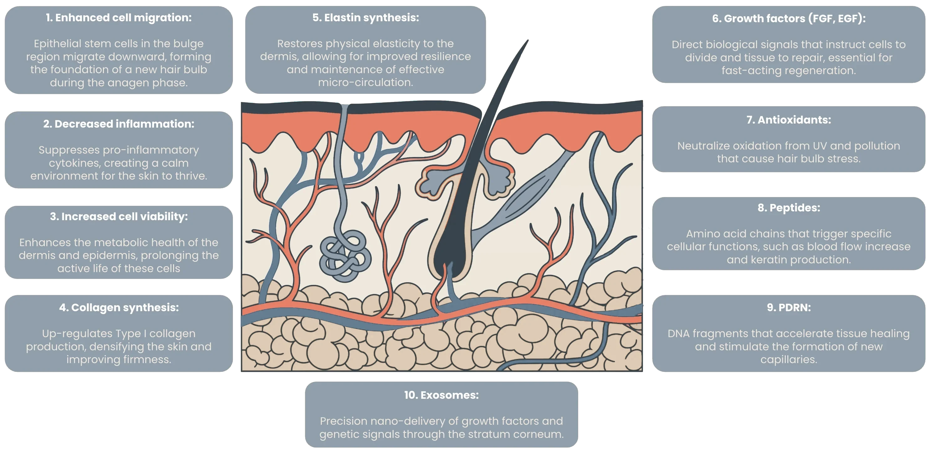
Exosome characterization
NTA confirmed a native exosome fraction, with vesicle identity confirmed by CD63 and syntenin/SDCBP markers. We disclose the count with its method, because a particle count is only meaningful alongside the instrument used.
NTA · average 154.1 nm · range 30–200 nm · 5.5 × 10⁹ particles/mL
Representative result. Full statistics (n, error, test) in the technical dossier.
Concentration (NTA)
Average diameter (NTA)
Measured size range
Nanoparticle tracking analysis (NTA). Vesicle identity confirmed by CD63 and syntenin/SDCBP markers.
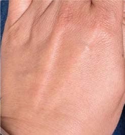

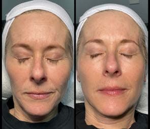
Additional clinical metrics, including before-and-after photos and quantified treatment metrics, are available with MCC's technical documentation.
Safety
Not derived from human tissue
Clean, contaminant-free production without human-derived inputs.
Heavy metals
Rigorously tested for mercury, arsenic, and cadmium.
Allergens
MCC is salmon-derived and contains salmon origin proteins. Individuals who are allergic to salmon should consult a medical professional and patch-test prior to use. Finished products may contain fish allergens. Third-party RIPT is non-reactive; documentation available upon request.
Physiologically-tuned, not spiked
Growth factors are present at the concentrations living cells naturally produce, not super-physiologic doses of single factors. No cytotoxicity observed in vitro.
The cell culture process underlying the company's cultivated salmon has also undergone extensive safety testing. As with any cosmetic ingredient, the potential for allergenicity exists; patch testing is recommended for sensitive skin.
- [1]
Squadrito F, Bitto A, Irrera N, et al. Pharmacological Activity and Clinical Use of PDRN. Front Pharmacol. 2017;8:224.— Adenosine A2A-receptor mechanism (polynucleotides). PubMed ↗
- [2]
Colombo M, Raposo G, Théry C. Biogenesis, secretion, and intercellular interactions of exosomes and other extracellular vesicles. Annu Rev Cell Dev Biol. 2014;30:255–289.— Exosome / EV biology. PubMed ↗
- [3]
Théry C, Witwer KW, Aikawa E, et al. Minimal information for studies of extracellular vesicles 2018 (MISEV2018). J Extracell Vesicles. 2018;7(1):1535750.— EV characterization standards. PubMed ↗
- [4]
Mehta RC, Fitzpatrick RE. Endogenous growth factors as cosmeceuticals. Dermatol Ther. 2007;20(5):350–359.— Growth factors in skin. PubMed ↗
- [5]
Alquraisy A, Wilar G, Suhandi C, et al. A Comprehensive Review of Stem Cell Conditioned Media Role for Anti-Aging on Skin. Stem Cells Cloning. 2024;17:5–19.— Conditioned-media skin anti-aging — review. PubMed ↗
- [6]
Naughton GK, Jiang LI, Makino ET, et al. Targeting Multiple Hallmarks of Skin Aging: Efficacy of a Growth Factor–Based Skin Care Serum. Dermatol Ther (Heidelb). 2022;13(1):169–186.— Fibroblast conditioned-media serum; clinical skin rejuvenation. PubMed ↗
- [7]
Kwon TR, Oh CT, Choi EJ, et al. Conditioned medium from human bone-marrow MSCs promotes skin moisturization and effacement of wrinkles in UVB-irradiated mice. Photodermatol Photoimmunol Photomed. 2015;32(3):120–128.— Pro-collagen synthesis; anti-wrinkle. PubMed ↗
- [8]
Noh CH, Park S, Seong HR, et al. An Exosome-Rich Conditioned Medium from Human Amniotic Membrane Stem Cells Facilitates Wound Healing via Reepithelialization, Collagen Synthesis, and Angiogenesis. Cells. 2023;12(23):2698.— Exosome-rich CM; collagen + angiogenesis. PubMed ↗
- [9]
Chien WY, Huang HM, Kang YN, et al. Stem cell-derived conditioned medium for alopecia: a systematic review and meta-analysis. J Plast Reconstr Aesthet Surg. 2023;88:182–192.— Hair density & thickness — meta-analysis. PubMed ↗
- [10]
Shin H, Won CH, Chung WK, Park BS. Clinical Trials of Hair Regeneration Using Conditioned Media of Adipose-Derived Stem Cells in Pattern Hair Loss. Curr Stem Cell Res Ther. 2017;12(7):524–530.— Conditioned media for pattern hair loss. PubMed ↗
- [11]
Ahangar P, Mills SJ, Smith LE, et al. Human gingival fibroblast secretome accelerates wound healing through anti-inflammatory and pro-angiogenic mechanisms. NPJ Regen Med. 2020;5:24.— Fibroblast secretome; reduced scarring. PubMed ↗
- [12]
Kuncorojakti S, Pratama AZA, Antujala CA, et al. Adipose mesenchymal stem cell secretome accelerates healing of cutaneous burn wounds. Vet World. 2024;17(7):1545–1554.— Secretome; collagen density + angiogenesis. PubMed ↗
- [13]
Nifontova G, Safaryan S, Khristidis Y, et al. Advancing wound healing by hydrogel-based dressings loaded with cell-conditioned medium: a systematic review. Stem Cell Res Ther. 2024;15(1):371.— Conditioned-media wound healing — review. PubMed ↗
References above describe conditioned media / cellular complexes and relevant molecules present in MCC – they are studies of other conditioned media and the underlying biology, not of MCC itself. MCC-specific study summaries are available in the technical dossier on request.
Interactive · MCC Explorer
Ask the dataset directly.
Query the proteomics, composition, formulation, and characterization data in plain language. Answers are synthesized from verified, high-fidelity datasets. MCC Explorer is AI-guided and can occasionally make mistakes. Primary data will be openly shared upon request to corroborate all findings.
Query the proteins, peptides, biological activity, composition, and technical data for Marine Cellular Complex. Select a topic or type your own question.
- INCI
- Salmon Mesenchymal Cell Conditioned Media
- Form / appearance
- Aqueous media, mild pink, clear liquid without visible particles.
- Odor
- Neutral
- pH
- 6.5–7.5
- Osmolality
- 300–385 mOsm/kg
- Phase matching
- Keep oil & water phases within ±3 °C before combining
- Preservation
- No preservatives incompatibilities observed to date
- Storage
- Up to 12 months at −20 °C; avoid repeated freeze-thaw cycles.
- Formulation notes
- Cold process preferred to maximize bioactivity
- Bacterial endotoxin
- <0.500 EU/mL (limit ≤5.00 EU/mL)
- Total aerobic count
- <10 CFU/g (limit ≤10,000 CFU/g)
- E. coli
- Negative (not detected)
- Heavy metals (As/Cd/Hg/Pb)
- All below instrument detection limit
- Manufacturing
- cGMP · US FDA-registered facility (Wildtype, San Francisco)